

Contact us
E.mail : infos@eyecare.com
Address : 1 Dmshek,Maadi, Cairo
Eye Care : Sat. - Thu. 9:00am - 10:00pm

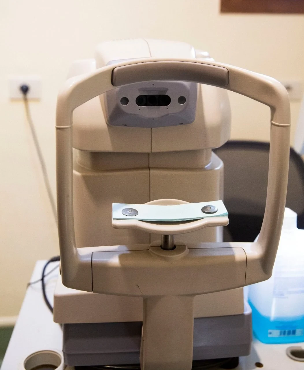
Specular microscopy
The Cornea & Corneal Endothelial Cell Examination
The cornea is one of the essential structures of the eye. It is the transparent, dome-shaped tissue covering the front of the eye. It protects the eye and plays a major role in focusing incoming light, contributing to clear vision.
The cornea consists of several specialized layers, each with an important function in maintaining its structure, transparency, and optical performance.
One of its most important components is the corneal endothelium, a single layer of cells lining the inner surface of the cornea. These cells help regulate fluid balance within the corneal tissue and maintain its transparency.
The Importance of Corneal Endothelial Cells
Corneal endothelial cells are located on the innermost layer of the cornea. They regulate the amount of fluid within the corneal tissue through their pump and barrier functions, helping maintain corneal clarity.
These cells have a limited ability to regenerate. Significant cell loss may affect their function, potentially leading to corneal swelling, reduced transparency, and impaired vision.
Endothelial cell density varies between individuals and may be affected by age, certain corneal diseases, and previous eye surgeries.
Specular Microscopy
At Eye Care Center, corneal endothelial cells are examined using the Topcon Specular Microscope SP-3000P. This diagnostic examination provides images of the endothelial cells and helps assess their density, size, and shape.
The examination provides several important measurements, including:
-
Endothelial Cell Density: The number of endothelial cells per square millimeter.
-
Polymegathism: Assessment of variations in cell size.
-
Pleomorphism: Assessment of cell shape and the proportion of hexagonal cells.
-
Overall Endothelial Cell Status: Evaluation of the general condition of the corneal endothelial layer.
When Is Specular Microscopy Needed?
An ophthalmologist may recommend this examination in several situations, including:
-
Certain inherited and acquired corneal diseases affecting endothelial cells.
-
Before cataract surgery, particularly in patients with risk factors that may affect corneal endothelial health.
-
Before secondary intraocular lens implantation, depending on the patient's condition.
-
When endothelial cell dysfunction or reduced endothelial function is suspected.
-
To monitor corneal health in selected patients following eye surgery or with conditions affecting the inner corneal layer.
Specular microscopy provides valuable information about corneal endothelial health and helps the ophthalmologist assess the cornea's suitability for certain surgical procedures and determine the most appropriate treatment plan.


Connect Us