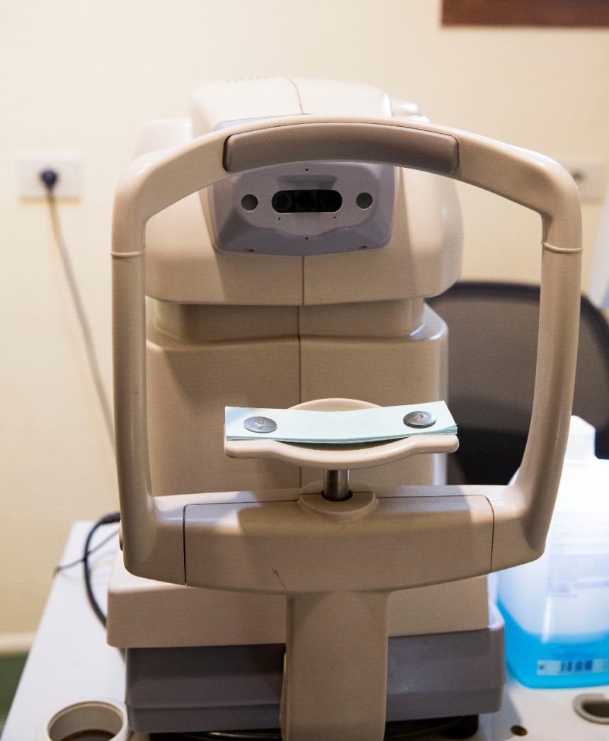
Specular microscopy
Specular microscopy – Topcon Specular Microscope SP-3000P : used to count the cells on the inner surface of the cornea, its important in some hereditary conditions and before cataract surgery and before and after ICL implantation (secondary lens inside the eye)
It is a diagnostic modality for imaging the corneal endothelium that allows for direct observation of the endothelial cell morphological characteristics either in a clinic or eye bank setting. Endothelial imaging using a specular microscope is routinely used in the assessment of endothelial health in various endothelial diseases, evaluation of the donor cornea prior to keratoplasty and postoperative follow-up after keratoplasty. A PubMed search was done using the keywords specular microscopy, corneal endothelium and endothelial imaging and appropriate references were included in the citation.
It is a simple and valuable tool for the evaluation of corneal endothelium in normal and diseased eyes. It is critical to perform the specular microscopy as per the standard guidelines and interpreted with a clear understanding of its limitations in various situations. Clinico-specular correlation helps in making an accurate diagnosis of a condition, assessing the functional reserve of the cornea and pre-operative evaluation in surgical management.

