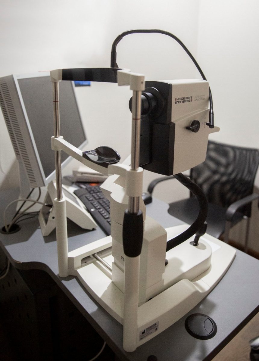
Fundus Fluorescein Angiography
Using the Heidelberg spectralis high detailed image of the retinal vasculature and to detect any abnormality in the circulation with special importance in some retinal diseases specially diabetis mellitus.
FFA – Technique
- Baseline color and black and white red-free filtered images are taken prior to injection. The black and white images are filtered red-free (a green filter) to increase contrast and often gives a better image of the fundus than the color image.
- A 6-second bolus injection of 2-5 cc of sodium fluorescein into a vein in the arm or hand
- A series of black-and-white or digital photographs are taken of the retina before and after the fluorescein reaches the retinal circulation (approximately 10 seconds after injection). The early images allow for the recognition of autofluorescence of the retinal tissues. Photos are taken approximately once every second for about 20 seconds, then less often. A delayed image is obtained at 5 and 10 minutes. Some doctors like to see a 15-minute image as well.
- A filter is placed in the camera so only the fluorescent, yellow-green light (530 nm) is recorded. The camera may however pick up signals from pseudofluorescence or autofluorescence. In pseudofluorescence, non-fluorescent light is imaged. This occurs when blue light reflected from the retina passes through the filter. This is generally a problem with older filters, and annual replacement of these filters is recommended. In autofluorescence, fluorescence from the eye occurs without injection of the dye. This may be seen with optic nerve head drusen, astrocytic hamartoma, or calcific scarring.
- Black-and-white photos give better contrast than color photos, which aren’t necessary because the filter transmits only one color of light.

