Eye Structures – Eyes are the first organs of the visual system, they are responsible for the ability to receive and process visual detail, they detect light and convert it into electro-chemical impulses in neurons which are transmitted to the visual centers in the brain.
In a number of ways, the human eye works much like a digital camera:
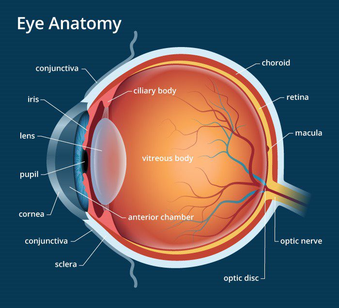
However in a little bit more detailed words about eye structures the eye consists of:
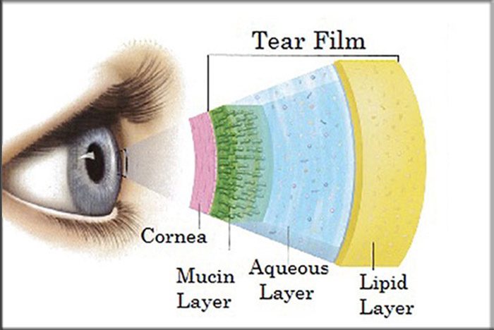
1. Tear film Layer
The Tear Layer (A component of the Lacrimal System) is the first layer of the eye that light strikes. It is clear, moist, and salty. It’s a very important layer and the main purpose is to keep the eye smooth and moist.
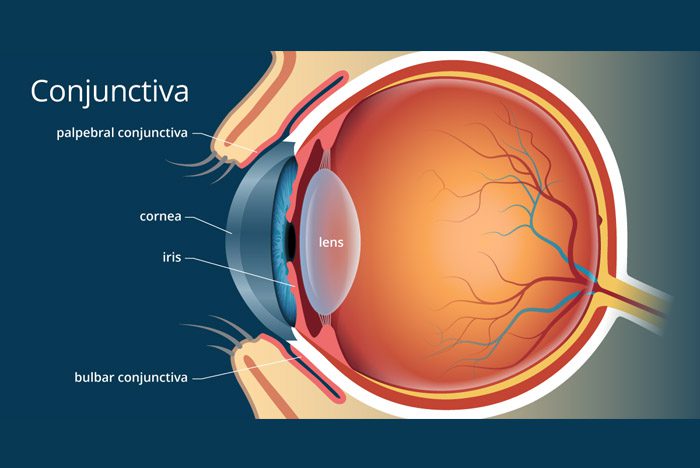
2. Conjunctiva
The conjunctiva is the clear, thin membrane that covers part of the front surface of the eye and the inner surface of the eyelids. It has two segments:
Bulbar conjunctiva. This portion of the conjunctiva covers the anterior part of the sclera (the “white” of the eye). The bulbar conjunctiva stops at the junction between the sclera and cornea; it does not cover the cornea.
Palpebral conjunctiva. This portion covers the inner surface of both the upper and lower eyelids. (Another term for the palpebral conjunctiva is tarsal conjunctiva.) The bulbar and palpebral conjunctivais continuous (see illustration). This feature makes it impossible for a contact lens (or anything else) to get lost behind your eye.
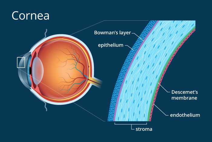
3. Cornea
The Cornea is the second structure that light strikes. It is the clear, transparent front part of the eye that covers the iris, pupil and anterior chamber and provides most of an eye’s optical power (in simple words: mostly if too flat = hyperopia/farsightedness; if too steep=myopia/nearsightedness). It needs to be smooth, round, clear, and tough. It is like a protective window. The function of the cornea is to let light rays enter the eye and converge the light rays.
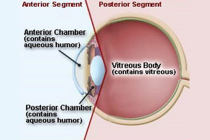
4. Anterior Chamber
The Anterior Chamber is filled with Aqueous Humor. Aqueous Humor is a clear, watery fluid that fills the space between the back surface of the cornea and the front surface of the vitreous, bathing the lens (The anterior and posterior chambers. Both are located in the front part of the eye, in front of the lens). The eye receives oxygen through the aqueous. Its function is to nourish the cornea, iris, and lens by carrying nutrients, it removes waste products excreted from the lens, and maintain intraocular pressure and thus maintains the shape of the eye. This gives the eye its shape. It must be clear to function properly.

5. Iris
The iris is the pigmented tissue lying behind the cornea that gives color to the eye and controls the amount of light entering the eye by varying the size of the pupillary opening. It functions like a camera. The color of the iris affects how much light gets in. The iris controls light constantly, adapts to lighting changes, and is responsible for near point reading (to see close, pupils must constrict)

6. Pupil
It is a variable-sized black circular opening in the center of the iris that regulates the amount of light that enters the eye. The pupil needs to be round in order to constrict.
A constricted pupil occurs when the pupil size is reduced to constriction of the iris or relaxation of the iris dilator muscle. The iris constricts with bright illumination, with certain drugs, and can be a consequence of ocular inflammation.
A dilated pupil is an enlarged pupil, resulting from contraction of the dilator muscle or relaxation of the iris sphincter. It occurs normally in dim illumination, or may be produced by certain drugs (mydriatics) or result from blunt trauma.
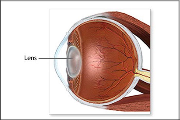
7. Lens
The lens is the natural lens of the eye (chrystaline lens). Transparent, biconvex intraocular tissue that helps bring rays of light to focus on the retina (It bends light, but not as much as the cornea). Suspended by fine ligaments (zonules) attached between ciliary processes. It has to be clear, has to have a power of about +16, and has to be pliable so it can control refraction (This becomes less pliable as you age leading to presbiopia).
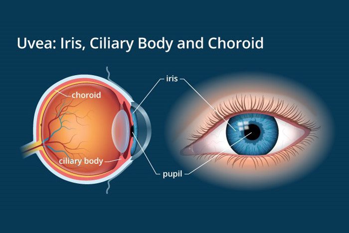
8. Ciliary Body
The circumferential tissue (a ring of tissue between the end of the choroids and the beginning of the iris) inside the eye composed of the ciliary muscle (involved in lens accommodation and control of intraocular pressure and thus the shape of the lens) and 70 ciliary processes that produce aqueous fluid.
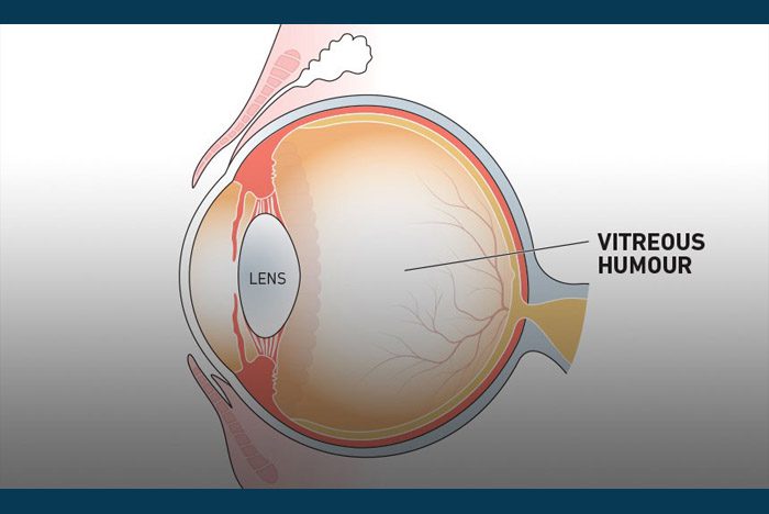
9. The Vitreous Humour.
Vitreous Humour is the transparent, colorless gelatinous mass that fills rear two-thirds of the eyeball, between the lens and the retina. It has to be clear so light can pass through it and it has to be there or eye would collapse.
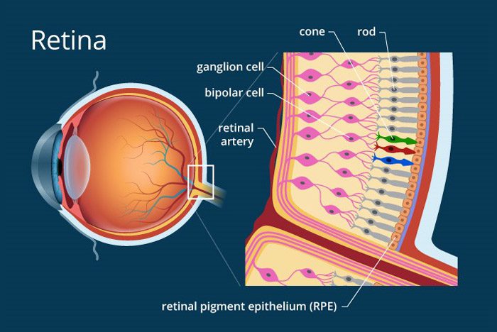
10. The Retina
The retina is the light-sensitive nerve tissue in the eye that converts images from the eye’s optical system into electrical impulses that are sent along the optic nerve to the brain, to interpret as vision. Forms a thin membranous lining of the rear two-thirds of the globe; consists of layers that include two types of cells: rods and cones. There is no retina over the optic nerve which causes a blind spot (This is the sightless area within the visual field of a normal eye. It is caused by an absence of light-sensitive photoreceptors where the optic nerve enters the eye.)
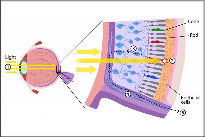
The cones are the light-sensitive retinal receptor cell that provides the sharp visual acuity (detail vision) and color discrimination; most numerous in macular area. Function under bright lighting.
The Rods are the light-sensitive, specialized retinal receptor cell that works at low light levels (night vision). The rods function with movement and provide light/dark contrast. It makes up peripheral vision.
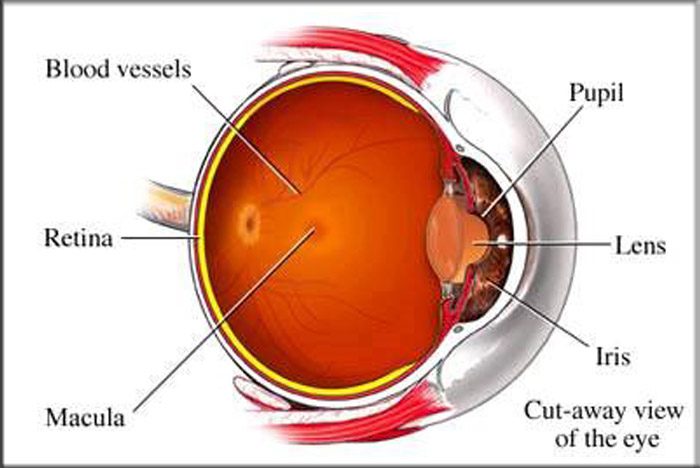
The Macula is the “yellow spot” in the small (3 °) central area of the retina surrounding the fovea. It is the area of acute central vision (used for reading and discriminating fine detail and color). Within this area is the largest concentration of cones.

The Fovea is the central pit in the macula that produces the sharpest vision. It contains a high concentration of cones within the macula and no retinal blood vessels.
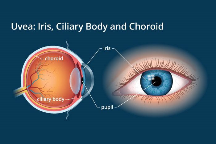
11. The Choroid
The vascular (major blood vessel), The central layer of the eye lying between the retina and sclera. Its function is to provide nourishment to the outer layers of the retina through blood vessels. It is part of the uveal tract.
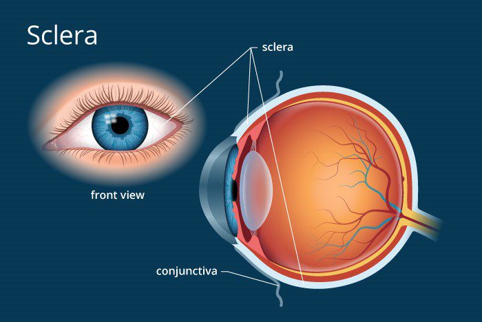
12. The Sclera
The sclera is the opaque, fibrous, tough, protective outer layer of the eye (“white of the eye”) that is directly continuous with the cornea in front and with the sheath covering the optic nerve behind. The sclera provides protection and form
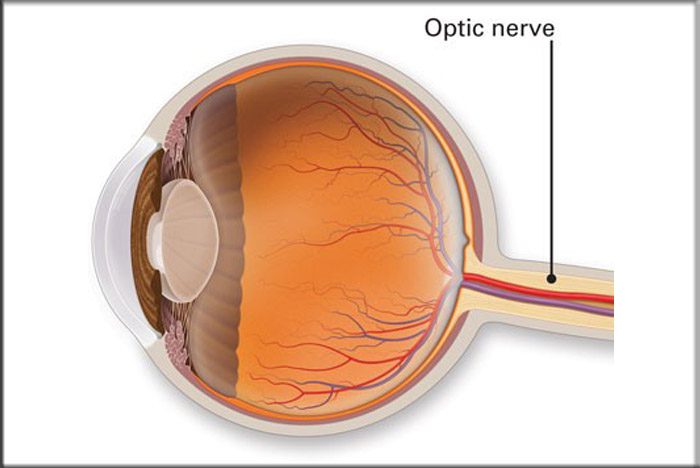
13. The Optic Nerve
The Optic Nerve is the largest sensory nerve of the eye. It carries impulses for sight from the retina to the brain. Composed of retinal nerve fibers that exit the eyeball through the optic disc, traverse the orbit, pass through the optic foramen into the cranial cavity, where they meet fibers from the other optic nerve at the optic chiasm.
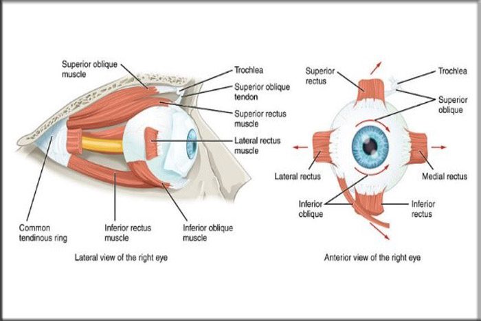
14. The Extra Ocular Muscles
There are six extraocular muscles in each eye in the simplest words their functions are:
Recti Muscles. There are four Rectus muscles that are responsible for straight movements: Superior (upward), Inferior (lower), Lateral (toward the outside, or away from the nose), and Medial (toward the inside, or toward the nose).
Oblique Muscles. There are two Oblique muscles that are responsible for angled movements. The superior oblique muscles control angled movements upward toward the right or left. Inferior oblique muscles control angled movements downward toward the right or left.


