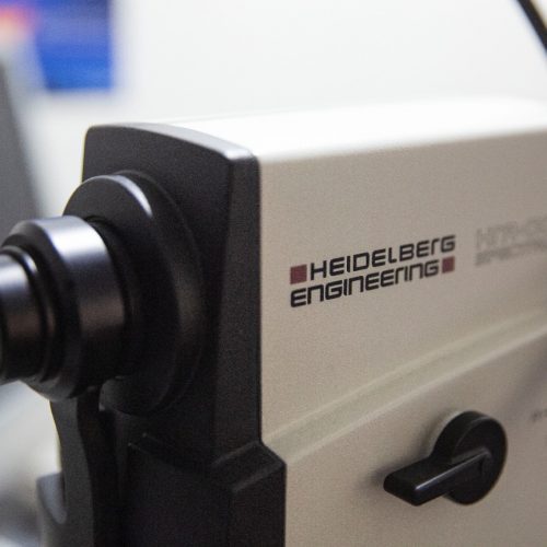
Fundus Fluorescein Angiography Using the Heidelberg spectralis high detailed image of the retinal vasculature and to detect any abnormality in the circulation with special importance in some retinal diseases specially diabetis mellitus. FFA – Technique Baseline color and black and white red-free filtered images are taken prior to injection. The black and white images are...
Category: <span>Eye Scan</span>
OCT – Optical Coherence Tomography
OCT – Optical Coherence Tomography Optical Coherence Tomography The Heidelberg spectralis OCT is one of the most advanced and precise equipment used to do the optical coherence tomograghy investigation giving high resolution images and precise information used in obtaining: MultiColor fundus photo Anterior Segment Scanning Nerve Fiber Layer Analysis Macula analysis OCT Angiography With non...
Dr. Fady Refaat
Dr. Naglaa Hassan
Professor of ophthalmology Memorial Institute of ophthalmology research MIOR Consultant of Ophthalmic imaging and investigations MIOR MD Cairo University
Dr. Kareem Adly
Visual Field Test
Visual Field Test Visual Field Test – An automated test to detect the visual field of the patient in a reproducible and internationally recognized format. This test is an eye examination that can detect dysfunction in central and peripheral vision which may be caused by various medical conditions such as glaucoma, stroke, pituitary disease, brain...
Cataract Biometry
Cataract Biometry Cataract Biometry – To measure the intra ocular lens power before cataract extraction surgery. Done by the Zeiss IOL Master device that uses a laser beam to precisely measure the eye structures and calculate the needed IOL for each patient very accurately also the A-scan Biometry is available which may be needed in...
Corneal Topography
Corneal Topography corneal topography – Using the Allegro Topolyser which uses Placido’s disc to optain a high resolution image og the corneal curvatures it is used in some cases of custom LASIK specially with high astigmatism or irregular corneal surface. Uses Corneal topography and tomography is most commonly used for the following purposes Refractive surgery:...
- 1
- 2


